Comparison of MRI slices at the mid calf showing the anatomy with an IP


XCR1+ conventional dendritic cell-induced CD4+ T helper 1 cell activation exacerbates cardiac remodeling after ischemic myocardial injury - Journal of Molecular and Cellular Cardiology

Three-dimensional reconstruction of the calf from the whole slice
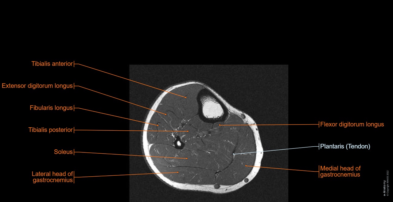
Lower limb: MRI anatomical atlas
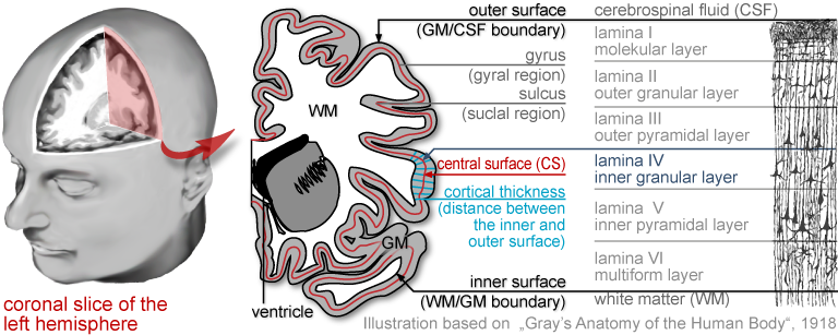
CAT12 Manual

Comparison of MRI slices at the mid calf showing the anatomy with an IP

Existing intravascular imaging technology for plaque characterization

Jean-Patrick BENIGNI, Medical Doctor, Polytech Paris-UPMC, Paris, polytech paris, Département de pédagogie

Presence of a valve at the saphenofemoral junction.
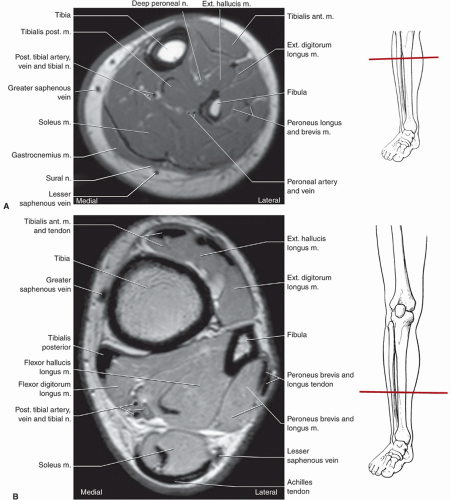
Foot, Ankle, and Calf

Dynamic study of the finger interphalangeal joint volar plate—motion analysis with magnetic resonance cinematography and histologic comparison

Gulf View Medical Centre - Did you know MRI's are commonly used to examine the brain, spine, joints, abdomen, and pelvis? Below are some imaging facts and information to consider: • An

PDF) Relationship between medical compression and intramuscular pressure as an explanation of a compression paradox

A comparison of peripheral imaging technologies for bone and muscle quantification: a technical review of image acquisition. - Abstract - Europe PMC
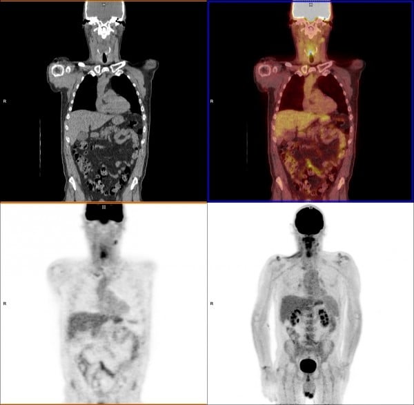
PET Scans & Imaging 101 Imaging Technology News