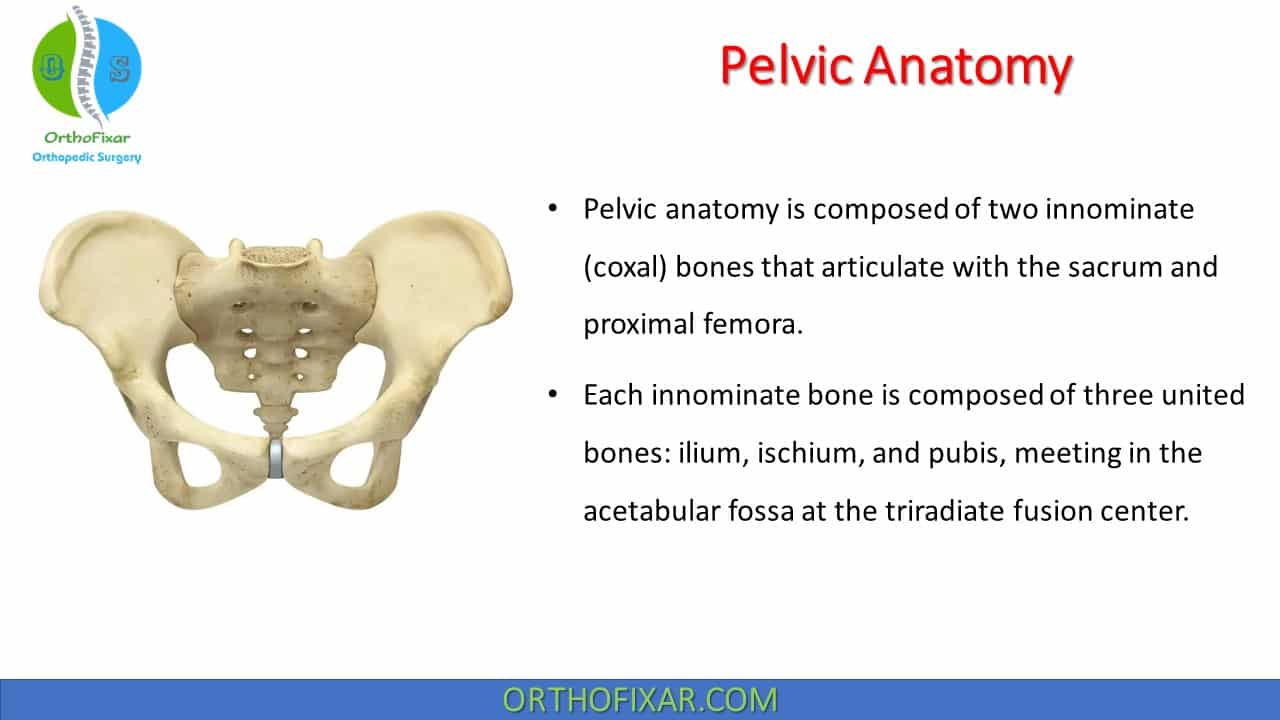
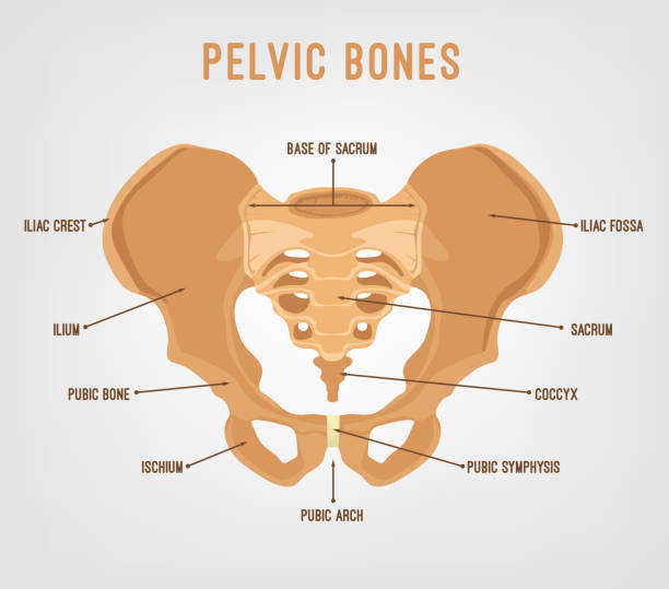
Figure showing the anatomy of the pelvic bone (A), the pelvic

Judet-Letournel classification of acetabular fractures into five

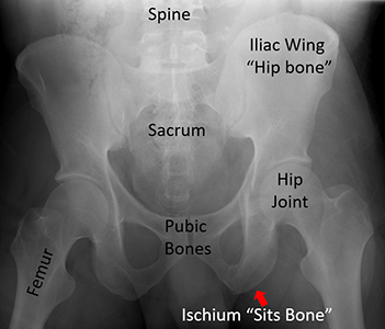
Example plain radiographs showing the Kellgren and Lawrence

Figure showing the anatomy of the pelvic bone (A), the pelvic

Robel Kebede GEBRE, Senior Postdoc Research Fellow, Doctor of Philosophy (PhD), Mayo Clinic - Rochester, Rochester
Anatomy, Bony Pelvis and Lower Limb, Hip - StatPearls - NCBI Bookshelf

Preoperative (A) and postoperative 1 st week (B) radiographs of the

Pelvic slices realignment. (a) shows the creation of a vertical

Preoperative (A) and postoperative 1 st week (B) radiographs of the

Example plain radiographs showing the Kellgren and Lawrence